

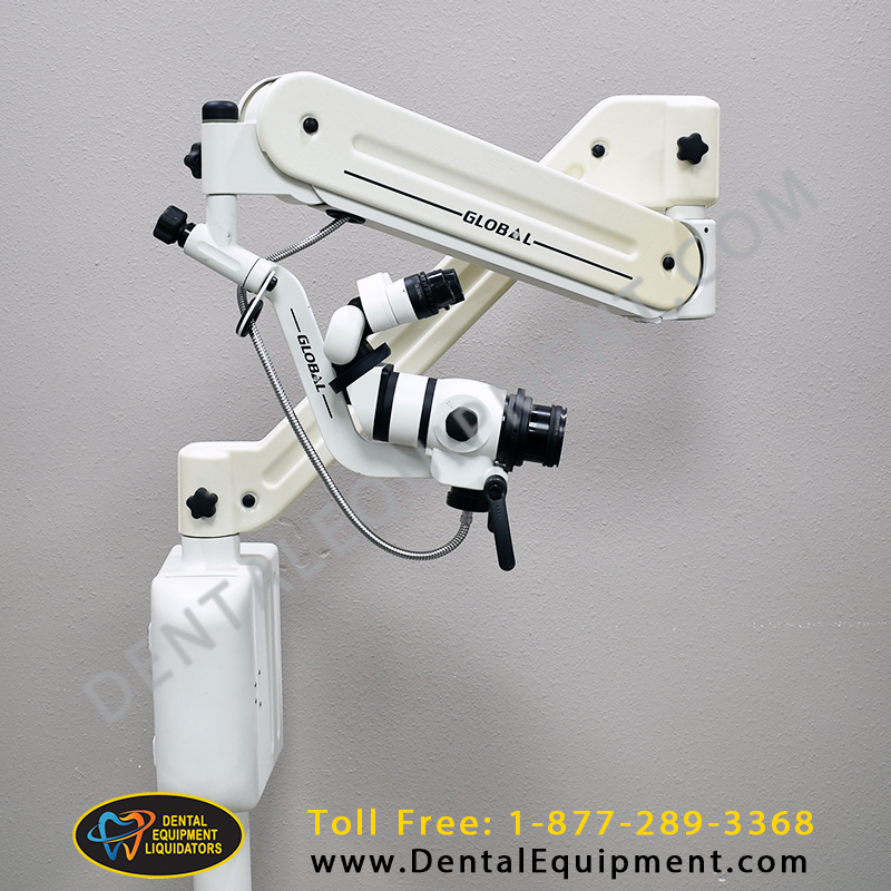
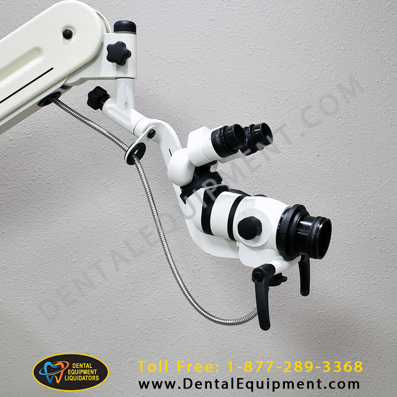
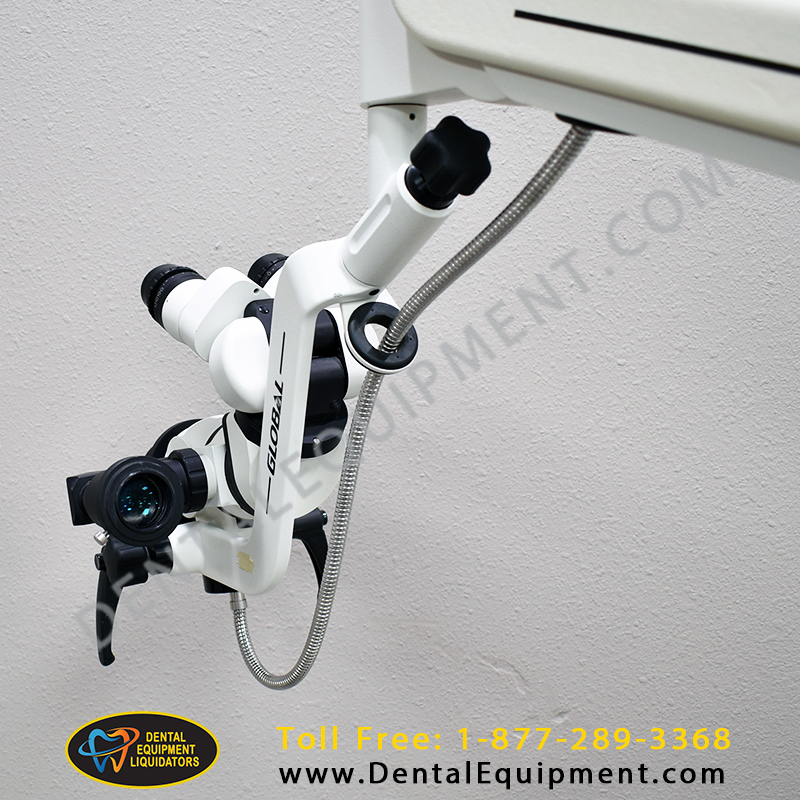
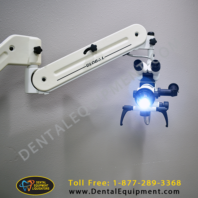
Microscope Global Surgical Lighted Microscope Mobile *Preowned*
Stock: 1
Warranty: 1 Year Parts, 90 Days Labor
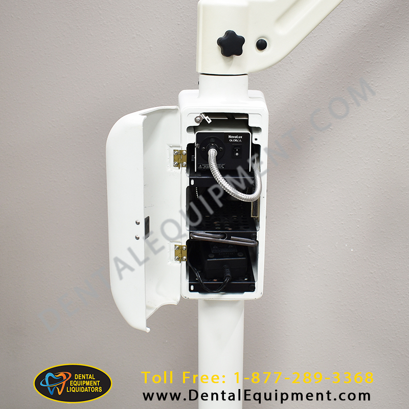
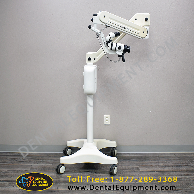
Global G3 Lighted Surgical Microscope with NovaLux Global Light Source
Applications: Endodontics, restorative dentistry, general dentistry, periodontal dentistry
6 Stage Magnification: 0.8, 0.5, .33, 1.25, 2.0, 3.0
Global Nova Lux Light Source: Halogen M1028G-250 is a 250mm focal length objective lens with fine-focus adjustment.
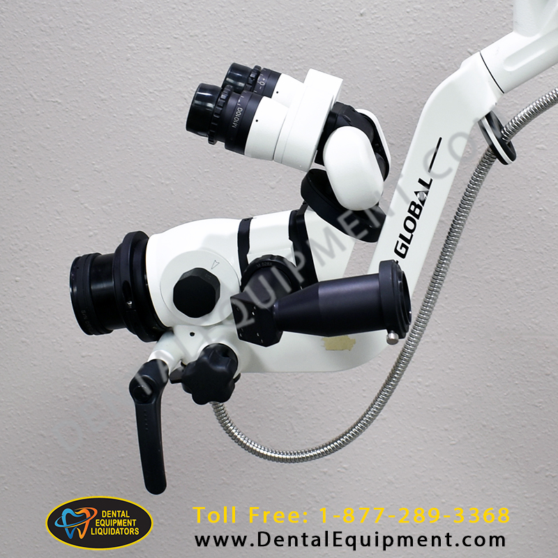
Global M1010G Eyepiece has adjustable optical focus/diopter settings with rubber eye bezels
Global M528X XMount Digital Camera Adapter connects consumer digital point-and-shoot cameras, camcorders, or DSLRs
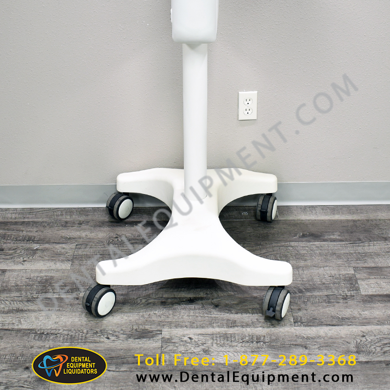
Global W725F Mobile Stand Professional-grade, mobile floor support system
Global Microscope offers many benefits for general practitioners, endodontists, and periodontists, based on its quality design and performance
The Floorstand Model is designed for portability around the office; It uses a compact H-base with four large casters and occupies minimal floor space. All four casters may be locked to prevent rolling.
Restorative Dentistry:
- Provides refinement in tooth and margin preparation.
- Allows for closer inspection of restorations and marginal tissues.
- Improved lighting and magnification aid in caries detection and removal.
- Improves detection and evaluation of coronal and root fractures and abnormalities.
- Facilitates cord placement for gingival retraction.
- Provides for better inspection of impressions.
- Helps with inspection of marginal fit of restoration (crowns, veneers, inlay/onlay, amalgam, composite).
- Facilitates in finishing and polishing of margins. Assists in gingival contouring or reshaping around teeth and implants.
- Assists in evaluation after cementation.
Benefits in the Dental Laboratory
- Assists in master die trimming
- Facilitates the location of bubbles, imperfections, and undercuts in the master die.
- Facilitates the marginal adaptation of wax or ceramic.
- Inspection of the internal aspect of metal casting or ceramic core for imperfections.
- Adaptation of restoration to die for the finest marginal integrity.
BENEFITS FOR ENDODONTISTS
The Global Dental Operating Microscope provides improved illumination and magnification, enhancing the precision and manipulation of all diagnostic and technical aspects of Endodontics.
- Diagnosis: improves visualization of fracture patterns, both of the coronal aspect of the tooth, the chamber, and into the root structure.
- Isolation, evaluation, and remediation of pulpal chamber obstructions, calcifications, and resorptions.
- Allowing for predictable pulp chamber visualization, the investigation of calcified anatomy can be conducted safer, with reduced loss of tooth structure, minimizing iatrogenic incidents.
- Three-dimensional visualization of root obstruction from separated instruments allowing for more successful removal of iatrogenic blockages.
- Endodontic retreatment cases successfully “disassembled” due to predictable removal of core build-ups; cemented posts, silver cones, and previous root fill materials.
- Careful evaluation of biomechanics allowing for more complete cleansing and disinfection of root canal systems.
- Thorough cleansing of deep caries can be completely illuminated, removed, and sealed from oral fluids.
- Manipulation of gingival tissue, for example, electrosurgical, suturing, and laser procedures can be more delicately directed with enhanced visualization.
- Endodontic surgical cases are enhanced as the complete lingual aspect of canal root structure can be more readily seen, allowing for complete root resection and retro fill procedures. Retrofill preparations can be directed to the proper alignment and depth. Enhanced surgical visualization allows for careful evaluation of suspected fractured root structure and periosseous defects.
The Global Dental Operating Microscope improves kinesiology during endodontic procedures. The clinician’s position when working with the scope allows for a more favorable upright working environment, helping to alleviate occupational neck, back and shoulder issues.
- Reduces operator fatigue and eyestrain during procedures.
- Provide a wonderful platform for photographic documentation.
BENEFITS FOR PERIODONTISTS
- Allows surgeon to minimize the size of the surgical site, reducing patient discomfort and healing time.
- Improves accuracy of microsurgical incisions and suturing with 6-0 through 8-0 sutures, permitting precise tissue/tissue and tissue/tooth approximation for primary would healing.
- Allows for better inspection and diagnosis of abnormal soft tissue lesions of the gingiva, palate, and mucosa.
- Improves visualization of root surface and adjacent intra-bony defects for definitive removal of calculus.
- Aids identification of micro-inflammation during re-evaluation following non-surgical therapy.
- Helps inspection of the quality of restorations and marginal tissues.
- Permits micro-level osseous surgery, facilitating bone removal without nicking the root surface and allows for better periodontal ligament preservation during ostectomy.
- Permits accurate subepithelial placement and suturing of membranes and subepithelial connective tissue grafts.
- Improves visualization of implant sites with minimal space between teeth and helps in evaluating the exact fit of implant prosthetic components and the health of marginal tissues around implants.
- Permits precise control of laser surgery on adjacent teeth without injury to root or implant surfaces.
- Permits accurate and easier root amputations and hemisection. Also helps with periapical surgeries when required during periodontal surgery.
- Facilitates sinus lift procedures through direct visualization of the sinus membrane during dissection.
- Permits fine dissection of the mandibular and mental nerves for lateral displacement during mandibular implant procedures.
- Permits location of the periodontal ligament for atraumatic elevation of roots and root tips during extraction with concurrent ridge preservation/augmentation.
- Improves detection and evaluation of root fractures and abnormalities.
- Provides upright working conditions, alleviating occupational neck, back, and shoulder problems.
- Provides high-resolution video and 35mm photography for patient education